Posts

Showing posts from 2013

Breast cancer

Demographics Breast cancer is the most common cancer among women and the second largest cause of cancer death. The incidence of breast cancer increases with age and peaks between the ages of 45-55 and plateaus from then onwards. The incidence of breast cancer is increasing due to increased screening.  Aeitiology and pathophysiology:  Breast cancer is caused by accumulation of genetic mutations leading to malignant growth.  Risk factors of breast cancer include:  Previous breast cancer Family history of breast cancer (if more than 2 first degree relatives have breast cancer, the risk to other members is doubled. )  Age Genetical - individuals with 4 relatively common genes are more susceptible to developing breast cancer. This includes BRCA 1, BRCA 2, CHEK2 and FGFR2. In individuals with these genes, lifetime risk of developing cancer may be up to 80% with a 60% risk of developing ovarian cancer.  Hormone replacement therapy with oest...

Headache Differentials

Image

Myeloproliferative disorders

Myeloproliferative disorders are diseases where one type of myeloid cell is produced excessively. See this link re what is the difference between myeloproliferative disorders and lukaemias? https://www.pathologystudent.com/?p=2153 Primary thrombocythaemia (essential thrombocythaemia) Involves clonal proliferation of the megakaryocyte line of cells leading to a raised platelet level.  Most commonly presents after 50 but may occur at any age. In the younger age groups, it is more common in women than men.  May present as abnormal thrombosis or bleeding, or may be asymptotic (up to 50%) and be found incidentally on routine blood tests. Neurological symptoms from microthrombosis (paraesthesia/burning pain) or signs from arterial /venous thrombosis.  Splenomegaly and/or hepatomegaly may be present in some patients Differentials : other haematological conditions may also caused a raised platelet count, and platelet count may be acutely raised secondary to some condi...

Occular melanoma

Image
Definition and demographics Occular melanomas are uncontrolled malignant proliferation of melanocytes in the eye. They can be divided into uveal melanomas, which arise in the iris, choroid body or ciliary body, and conjunctival melanomas which arise from the conjunctiva. Uveal melanomas are more common and is the most common type of primary eye malignancy, and second most common type of malignant melanoma.  Ocular melanoma is a rare condition which occurs equally in males and females and the with a peak incidence in people's 60s. It tends to affect fair skinned individuals more.  Aeitiology/pathophysiology Occular melanomas are caused by an accumulation of genetic changes. Sunlight exposure is a risk factor of developing melanoma. Other risk factors include being caucasion/fair skinned, having light coloured irides, melanosis or existing melanocytic naevi of the conjunctiva (moles), congenital ocular melanocytosis, neurofibromatosis and family history.  ...

colorectal cancer

Demographics:  Colon cancer is a common malignancy, it primarily presents in older patients, however it may occur earlier in two hereditary conditions which leads to colon cancer. Familial adenomatous polyposis (FAP) is a autosomnal dominant condition which causes the formation of multiple polyps in the colon. The condition tends to cause cancer in patients by the age of 40. Hereditary non-polyposis colorectal cancer (HNPCC) also causes colon cancer in younger patients. HNPCC is also associated with endometrial, gastric, renal, uteric and CNS cancers.  Risk factors and causes Crohn's and Ulcerative colitis increases colorectal cancer risk History of small bowel cancer, endometrial cancer, breast cancer or ovarian cancer Family history - family member with cancer before the age of 60 Specific hereditary causes : FAP and HNPCC Low fibre diet and high red meat diet is thought to increase risk Sedentary lifestyle, obesity, smoking, high alcohol intake Diabetes m...

Describing skin lesions

Image
Describing lesions  Site, Distribution and symmetry Type of lesion Size Shape - edges, well demarcated? regular/irregular shape? Surface -color and texture, scaly vs non-scaly (epidermis lesions are scaly) Special features - fluid filled?, mobility, temperature/inflammation, other features.

Glucocorticoids

Image
Today I made up a mnemonic for remembering the effects of excessive glucocorticoids:  "Grossly Fat Pigs Can Never Ice-skate Harmlessly" Cows however.... 

Pleural effusion

Image
Definition Pleural effusion is a collection of fluid in the pleural space between the visceral pleura surounding the lungs and the parietal pleural attatched to the throacic wall. Demographics Pleural effusions are relatively common, but it is a manifestation of other underlying diseases. The prevalence and incidence of pleural effusions is therefore dependent on it's cause. Pathophysiology/causes Pleural fluid is usually filtered from the parietal pleura into the pleural space, due to hydrostatic pressure from the capillaries, negative pressure in the pleural space and pleural oncotic pressure. The visceral pleura is perfused by the pulmonary system which has a lower pressure, and fluid in the pleural space is therefore drawn back in through the visceral pleura due to the higher oncotic pressure in plasma. Some of this fluid is then drained by the lymphatic system.

Chronic Obstructive Pulmonary Disease (COPD)

Image
Definition: Chronic obstructive pulmonary disease is defined as a condition of restricted airflow which is not fully reversible. It is often a progressive disease caused by abnormal inflammatory responses to obnoxious substances.  Demographics: COPD is a common condition, The prevalence of COPD increases with age, with most patients being diagnosed at around the age of 50.  Aeitiology and pathophysiology:  Airway obstruction in COPD is caused by;  1) increase in mucus secretion, leading to obstruction.  many COPD patients show increased number of goblet cells in their airways, which block airways.  2) inflammation and scarring to the airways, leading to narrowed airways.  Inflammatory cells infiltrate the bronchi and bronchiole walls. The inflammation causes scarring and thickening of walls which cause small airways to narrow. Initially, inflammation is reversible, but this become irreversible with continued smoking. As the dis...

Tumour markers

Tumour markers are molecules found in the blood which are sometimes secreted by some tumours. They may be helpful in confirming a diagnosis where cancer is suspected, but they are not by themselves diagnostic and may be present because of a number of other processes. Tumour markers are not routinely used as screening tests as they do not have high sensitivities at an early stage in the disease. In some cancers, tumour markers help monitor the effectiveness of the treatment, and tumour markers may be helpful in determining prognosis. Some tumour markers include: CA 27, CA29 - Breast cancer , also seen in colonic, gastric, hepatic, lung, pancreatic, ovarian and prostate cancer, other breast liver and kidney disorders and ovarian cysts. CEA - Colorectal cancer , also seen in lung, gastric, pancreatic, breast and bladder cancers, medullary thyroid, and other head and neck, cervical and hepatic cancers, lymphomas, melanomas, smoking, peptic ulcers, inflammatory bowel diseases, pancr...

Nausea and vomiting and antiemetics

Image
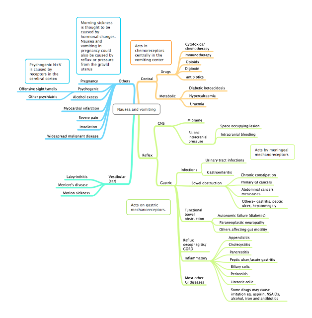
If a patient presents with nausea and vomiting, a clear cause should be established before perscribing antiemetics, as nausea and vomiting may be a sign of serious illness such as diabetic ketoacidosis, and may be reversed by treating the cause, for example in digoxin overdose. The cause of nausea and vomiting may be apparent with history and examination. patients with persistent nausea and vomiting should also be assessed for their fluid status, and for any electrolyte abnormalities caused by the vomiting. Causes of nausea and vomiting Nausea and vomiting may be caused centrally through stimulation of the vomiting center and the chemoreceptor trigger zone in the medulla, or caused afferent signals due to irritation in other areas of the body, usually in the gastrointestinal tract, but also due to disease in the central nervous system or the vestibular system in the ear. 

Lung Cancer

Lung cancer Definition : Uncontrolled invasive (malignant) growth originating from the lungs Presentation:   Depends on the site of the tumour and the type of cancer.  Patients may present with Chest signs and symptoms : (more common in cancers occurring centrally in the bronchial tree, late symptoms in peripheral cancers, may also present on a background of existing lung problems eg. COPD or emphysema)  *Haemoptysis *Cough Wheeze Stridor Pain Hoarse voice clubbing lymphadenopathy localized chest signs superior vena cava obstruction - caused by compression of the superior vena cava

Chemotherapy

Image
Malignancy occurs when cells mutate to replicate uncontrollably, outside of the body's normal control mechanisms and immune response, and gains the ability to invade into other tissues. Cell cycle As cancer cells tend to replicate faster than other cells, chemotherapy primarily affects cancer cells by affecting their replication.

Troponins and MI meds

causes of a raised troponin:  Myocardial infarction Massive pulmonary embolism - leads to increased work of the right ventricles which damage cardiac muscles releasing troponin Chronic kidney disease (especially in later stages, although they may also have concurrent acute coronary syndrome - look for an increase from baseline)  Myocarditis  Sepsis/other critically ill (increased work of the heart leading to myocyte damage)  Aortic dissection severe tachycardia/bradycardia Stroke or subarachnoid haemorrhage Medical treatment for patients with acute coronary syndromes on presenting (before PCI) :  M orphine O xygen N itrates A spirin C lopidigrel Medicines after an MI A torvastatin B- blocker C heck all else

Facial nerves

Image
5 main branches of the facial nerve: To Zanzibar By Motor Car Temporal Zygomatic Buccal Mandibular Cervical

Otitis Media with Effusion (Glue ear)

Image
Definition: Inflammation of the middle ear with the collection of fluid, without acute signs of inflammation. Also known as glue ear. Pathophysiology:  The pathophysiology of glue ear is not fully known, however it is thought to be associated with Eustachian tube dysfunction. This is either thought to lead to a sterile accumulation of fluids that predisposes the middle ear to infections, or is thought to cause more acute infections of the middle ear that then leads to chronic effusion. Children with cleft palate also often have glue ear as their tensor veli palatini muscle does not attatch properly to the soft palate such that the Eustachian tube does not open on swallowing or mouth opening.

Innervation of the vocal cords

Image
The vocal cords are innervated by the superior laryngeal nerve and the recurrent laryngeal nerves which are branches of the vagus nerve (cranial nerve X). The superior laryngeal nerve splits into the external and internal branches. The internal branch contains sensory nerves from the larynx whereas the external branch supplies the cricothyroid muscle which tenses the vocal cords. Damage to the superior laryngeal nerves can be caused by thyroid surgery. Unilateral damage to the superior laryngeal nerve causes slight voice changes, usually affecting pitch. Bilateral damage to the superior laryngeal nerves reduces the ability for the vocal cords to close increasing the risk of aspiration. The recurrent laryngeal nerves supply the other muscles of the vocal cords, including the posterior cricoarytenoid muscle which is the only muscle which opens the vocal cords. The recurrent laryngeal nerves have a different course on the left and the right. The left is longer and descends down to...

Community acquired pneumonia

Image
Definition:  Pneumonia is the inflammation of the terminal bronchioles and alveoli, characterised by consolidation of the affected part by inflammatory cells, exudate and fibrin. Pneumonia is most commonly caused by infection. Community acquired pneumonia is defined as pneumonia presenting in the community or in the first 2 days of hospital admission. Pneumonia occurring after this time is more likely to be hospital acquired and caused by a different set of mircoorganisms.

Diabetic Ketoacidosis (Adults)

Image
Definition : A major life threatening metabolic condition occurring in diabetic patients leading to hyperglycaemia , ketoacidosis , dehydration and ketonuria . Pathophysiology:  The lack of insulin primarily leads to hyperglycaemia and ketonaemia:  Insulin insufficiency leads to reduced uptake of glucose into cells and increased blood concentration of glucose.  As glucose are not taken into cells, alternative sources of energy are used.  Most cells break down fatty acids instead of glucose which forms ketones. Ketone levels therefore reflects the glucose deficit in cells.  Lack of insulin also leads to an increase in opposing hormones which lead to increased glycogenolysis, gluconeogenesis and lipolysis. 

Syncope differentials

Image
Definition: The transient loss of conciousness caused by transient global hypoperfusion of the brain. Syncope is sudden onset, occurs for a short duration, and is characterised by spontaneous complete recovery. Causes and Differentials:  Syncope presents as a transient loss of conciousness but is defined by its pathophysiology (transient global hypoperfusion) and is therefore different to other causes of loss of conciousness. Syncope can be divided into 3 main types which are summarised below:  Reflex syncope is the most common type.  

Notes on Pediatrics

Image
Patterns of childhood illnesses 

Quantitative chemistry

Image
Basic concepts Matter: anything which has mass and occupies space Atoms: the smallest chemical unit, indivisible by chemical means Elements: 1 type of atom which may or may not be bonded.  Compounds: Substances made from different types of atoms bonded Molecule: A covalently bonded unit.  Pure substances: A single type of compound or element Mixtures: 2 or more different types of compounds and/or elements, separable by physical means. Isotopes: Different types of atoms with the same number of protons but different number of neutrons. Isotopes may be radioactive.  1.1.1 Apply the mole concept to substances 1.1.2 Determine the number of particles and the amount of substance in moles. 

Cardiac Tamponade

Image
Definition: Accumulation of fluid in the pericarial space leading to restriction of the heart's pumping action and reduced cardiac output which may lead to hypoperfusion and failure of other organs. Different to cardiac effusion which is an umbrella term for a fluid collection in the pericardial space with or without haemodynamic compromise.  Pathophysiology A cardiac tamponade is caused by an increase in the pressure in the pericardial sac. The pericardial sac usually has a level of compliance (stretch) and therefore an increase in fluids in this space initially has little effect on the pressure. It is only when the sac has reached the limit of it's stretch that the pressure increases. The compliance of the pericardial sac can also be increased in a chronic effusion and as much of 2000mls of fluid can be present before it causes a tamponade. Compliance can also be reduced by some disease states.