Pleural effusion
Definition
Pleural effusion is a collection of fluid in the pleural space between the visceral pleura surounding the lungs and the parietal pleural attatched to the throacic wall.
Demographics
Pleural effusions are relatively common, but it is a manifestation of other underlying diseases. The prevalence and incidence of pleural effusions is therefore dependent on it's cause.
Pathophysiology/causes
Pleural fluid is usually filtered from the parietal pleura into the pleural space, due to hydrostatic pressure from the capillaries, negative pressure in the pleural space and pleural oncotic pressure. The visceral pleura is perfused by the pulmonary system which has a lower pressure, and fluid in the pleural space is therefore drawn back in through the visceral pleura due to the higher oncotic pressure in plasma. Some of this fluid is then drained by the lymphatic system.

A pleural effusion occurs when any of these factors are affected. This may be due to disease in the pleura, disease in the lung, or disease elsewhere which may affect capillary oncotic pressure or hydrostatic pressure.
Presentation
Pleural effusions present as dyspnoea associated with pleuritic chest pain.
Symtoms associated to the cause may also be present. risk factors related to underlying disease should be enquired about. eg. asbestos exposure, drugs, previous ischaemic heart disease etc.
Signs: pleural effusions present with decreased chest expansion on the side of the effusion, stony dullness to percussion, reduced breath sounds, and reduced tactile vocal fremitus. Sometimes bronchial breathing may be heard at the level of the fluid.
Differential diagnoses
Causes of pleural effusions can be divided into transudative causes or exudative causes. Transudates are caused by changes in hydrostatic or oncotic pressures, usually due to increase pulmonary venous pressure or hypoproteinaemia. On the other hand, exudates are caused by drawing in of water due to changes in capillary permeability or inflammatory processes (such as infection or malignancy). Exudates are are characterised by a high protein content (usually a protien level of over 35g/L is diagnostic of an exudative pleural effusion, and under 25g/L is diagnostic of a transudative cause. If protein levels is in between, light's criteria is used.
Transudative causes of pleural effusions include:
Exudate causes include
Investigations and management
Clinical history and examination and chest x ray is often adequate for determining whether the pleural effusion is caused by a transudate or an exudate.
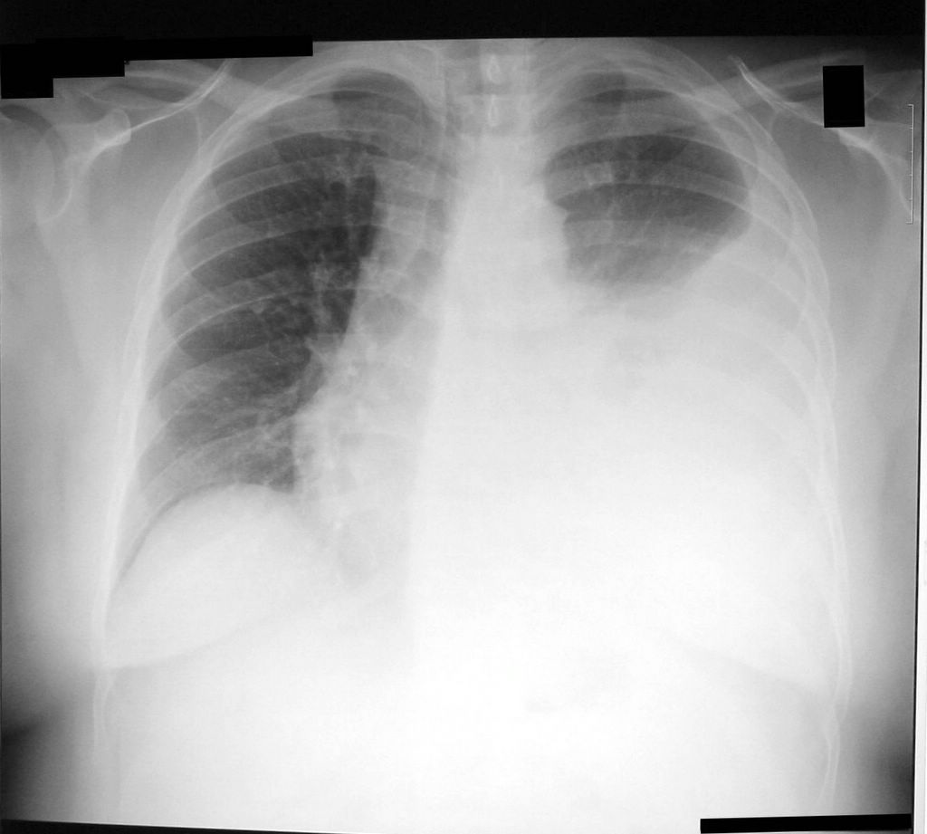 Chest x rays tend to show a whitened area in the lungs with a meniscus level, or may show loss of costophrenic angles. If the patient is supine pleural fluid may only cause a haziness on the affected side. Transudates tend to cause bilateral effusions.
Where an transudate cause is established, the cause should be treated, and a pleural fluid sample is not needed unless presentation is atypical or the patient does not respond to treatments.
Pleural fluid aspiration should be carried out where the effusion is thought to be caused by an exudate or where diagnosis is unclear clinically.
The fluid can be aspirated with a green needle into blood culture bottles and a sterile bottle and the following tests should be carried out:
protein and lactate dehydrogenase (used to determine if the pleural effusion is a transudate or exudate. If protein levels are between 25g/L and 35g/L, light's criteria should be used.
pH
Gram stain- parapneumonic effusions
AAFB stain
Cytology- may show malignant cells, lymphocycosis may suggest TB or malignancy, neutrophils suggest infection
Microbiological culture
pleural fluid may be
frankly bloody - do haematocrit of pleural fluid, malignancy, trauma, pulmonary embolus with infarction, benign asbestos, post cardiac injury syndrome
pus - empyema
Blood tinged- infection
Foul smelling - anaerobic infection
Milky - chylothroax, pseudochylothorax, - lymphoma and other malignancy, trauma, and other causes.
Other investigations:
Bloods: Liver function (albumin) FBC, CRP, amylase or lipase for pancreatitis, renal function
Imaging:
Ultrasound - good for ultrasound guided aspirations and may show fibrinous septations. Shows pleural thickening. May be used to position chest tubes or localise loculated effusions
CT - helpful in assessing underlying lung and differentiating between benign and malignant pleural thickening.
Invasive : percutaneous biopsy- if pleural malignancy or TB is suspected, thoracoscopy - if other test have failed. Bronchoscopy - clinical features of bronchial obstruction.
Further management depends on the diagnosis.

Chest drains and aspirations are useful in relieving symptoms, and prevention of recurrence can be achieved by using sclerosing agents. If chemical pleurodesis (forming adhesions between the 2 pleural layers) fail, surgical pleurodesis or pleurectomy may be considered.
From: SJ Bourke, Lecture notes - respiratory medicine, 7th ed. Oxford, Blackwell publishing, 2007
http://emedicine.medscape.com/article/299959-overview
http://thorax.bmj.com/content/58/suppl_2/ii8.full
Pleural effusion is a collection of fluid in the pleural space between the visceral pleura surounding the lungs and the parietal pleural attatched to the throacic wall.
Demographics
Pleural effusions are relatively common, but it is a manifestation of other underlying diseases. The prevalence and incidence of pleural effusions is therefore dependent on it's cause.
Pathophysiology/causes
Pleural fluid is usually filtered from the parietal pleura into the pleural space, due to hydrostatic pressure from the capillaries, negative pressure in the pleural space and pleural oncotic pressure. The visceral pleura is perfused by the pulmonary system which has a lower pressure, and fluid in the pleural space is therefore drawn back in through the visceral pleura due to the higher oncotic pressure in plasma. Some of this fluid is then drained by the lymphatic system.
A pleural effusion occurs when any of these factors are affected. This may be due to disease in the pleura, disease in the lung, or disease elsewhere which may affect capillary oncotic pressure or hydrostatic pressure.
Presentation
Pleural effusions present as dyspnoea associated with pleuritic chest pain.
Symtoms associated to the cause may also be present. risk factors related to underlying disease should be enquired about. eg. asbestos exposure, drugs, previous ischaemic heart disease etc.
Signs: pleural effusions present with decreased chest expansion on the side of the effusion, stony dullness to percussion, reduced breath sounds, and reduced tactile vocal fremitus. Sometimes bronchial breathing may be heard at the level of the fluid.
Differential diagnoses
Causes of pleural effusions can be divided into transudative causes or exudative causes. Transudates are caused by changes in hydrostatic or oncotic pressures, usually due to increase pulmonary venous pressure or hypoproteinaemia. On the other hand, exudates are caused by drawing in of water due to changes in capillary permeability or inflammatory processes (such as infection or malignancy). Exudates are are characterised by a high protein content (usually a protien level of over 35g/L is diagnostic of an exudative pleural effusion, and under 25g/L is diagnostic of a transudative cause. If protein levels is in between, light's criteria is used.
Transudative causes of pleural effusions include:
- Left ventricular failure
- Liver cirrhosis
- Hypoalbuminaemia
- Peritoneal dialysis
- Hypothyroidism
- Nephrotic syndrome
- Mitral stenosis
- Pulmonary embolism
- Constrictive pericarditis
- Urinothorax
- Superior vena cava obstruction
- Ovarian hyperstimulation
- Meigs' syndrome
Exudate causes include
- Malignancy (secondary met- lung, breast, ovarian, GI, and lymphoma, primary- mesothelioma)
- Parapneumonic effusions
- Pulmonary infarction
- Rheumatoid arthritis
- Autoimmune diseases
- Benign asbestos effusion
- pancreatitis
- Postmyocardial infaction syndrome
- yellow nail syndrome
- Drugs
- Fungal infections
Investigations and management
Clinical history and examination and chest x ray is often adequate for determining whether the pleural effusion is caused by a transudate or an exudate.

Where an transudate cause is established, the cause should be treated, and a pleural fluid sample is not needed unless presentation is atypical or the patient does not respond to treatments.
Pleural fluid aspiration should be carried out where the effusion is thought to be caused by an exudate or where diagnosis is unclear clinically.
The fluid can be aspirated with a green needle into blood culture bottles and a sterile bottle and the following tests should be carried out:
protein and lactate dehydrogenase (used to determine if the pleural effusion is a transudate or exudate. If protein levels are between 25g/L and 35g/L, light's criteria should be used.
pH
Gram stain- parapneumonic effusions
AAFB stain
Cytology- may show malignant cells, lymphocycosis may suggest TB or malignancy, neutrophils suggest infection
Microbiological culture
pleural fluid may be
frankly bloody - do haematocrit of pleural fluid, malignancy, trauma, pulmonary embolus with infarction, benign asbestos, post cardiac injury syndrome
pus - empyema
Blood tinged- infection
Foul smelling - anaerobic infection
Milky - chylothroax, pseudochylothorax, - lymphoma and other malignancy, trauma, and other causes.
Other investigations:
Bloods: Liver function (albumin) FBC, CRP, amylase or lipase for pancreatitis, renal function
Imaging:
Ultrasound - good for ultrasound guided aspirations and may show fibrinous septations. Shows pleural thickening. May be used to position chest tubes or localise loculated effusions
CT - helpful in assessing underlying lung and differentiating between benign and malignant pleural thickening.
Invasive : percutaneous biopsy- if pleural malignancy or TB is suspected, thoracoscopy - if other test have failed. Bronchoscopy - clinical features of bronchial obstruction.
Further management depends on the diagnosis.

Chest drains and aspirations are useful in relieving symptoms, and prevention of recurrence can be achieved by using sclerosing agents. If chemical pleurodesis (forming adhesions between the 2 pleural layers) fail, surgical pleurodesis or pleurectomy may be considered.
From: SJ Bourke, Lecture notes - respiratory medicine, 7th ed. Oxford, Blackwell publishing, 2007
http://emedicine.medscape.com/article/299959-overview
http://thorax.bmj.com/content/58/suppl_2/ii8.full


Comments
Post a Comment