Muscle contraction
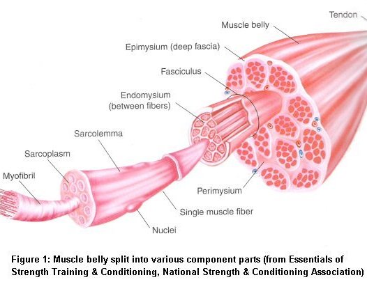
Skeletal muscles contain muscle fibres which span the whole length of the muscle. These are made up of subunits called sacromeres containing thick myosin filaments and thin actin filaments. Thick and thin filaments slide towards each other during contraction, the total effect of multiple sacromeres contracting leads to the shortening of the muscle.

Skeletal muscle contraction
- Acetyl choline is released at the neuromuscular junction. This causes the plasma membrane to depolarise.
- T-tubules which are continuous with the membrane depolarises.
- Depolarisation Activates dihydropyridine receptors and triggers calcium-release channels of the sarcoplasmic reticulum to open.
- Calcium enters the intracellular fluid and attaches to troponin.
- Troponin causes tropomyosin to change shape uncovering binding sites on actin.
- Myosin heads attatches to the exposed actin creating a cross bridge
- The myosin heads bends with energy stored from previously split ATP. this is called the power stroke.
- ADP and Pi are released.
- ATP attatches to an ATPase site on the myosin head which causes it to detach from the actin filaments.
- ATP splits causing the myosin head to bend back to its high energy state.
- Cycle repeats until Calcium is removed via a Calcium-ATPase pump in the sarcoplasmic reticulum. When calcium is removed, troponin returns tropomyosin to its original position preventing binding by myosin heads.

Myosin are made up of 2 protein subunits such that each molecule has 2 heads. During contraction, only one head can attach to actin at one time. This ensures that the filaments do not slide back to its original shape during contraction.


Comments
Post a Comment