Posts

Showing posts from October, 2013

Describing skin lesions

Image
Describing lesions  Site, Distribution and symmetry Type of lesion Size Shape - edges, well demarcated? regular/irregular shape? Surface -color and texture, scaly vs non-scaly (epidermis lesions are scaly) Special features - fluid filled?, mobility, temperature/inflammation, other features.

Glucocorticoids

Image
Today I made up a mnemonic for remembering the effects of excessive glucocorticoids:  "Grossly Fat Pigs Can Never Ice-skate Harmlessly" Cows however.... 

Pleural effusion

Image
Definition Pleural effusion is a collection of fluid in the pleural space between the visceral pleura surounding the lungs and the parietal pleural attatched to the throacic wall. Demographics Pleural effusions are relatively common, but it is a manifestation of other underlying diseases. The prevalence and incidence of pleural effusions is therefore dependent on it's cause. Pathophysiology/causes Pleural fluid is usually filtered from the parietal pleura into the pleural space, due to hydrostatic pressure from the capillaries, negative pressure in the pleural space and pleural oncotic pressure. The visceral pleura is perfused by the pulmonary system which has a lower pressure, and fluid in the pleural space is therefore drawn back in through the visceral pleura due to the higher oncotic pressure in plasma. Some of this fluid is then drained by the lymphatic system.

Chronic Obstructive Pulmonary Disease (COPD)

Image
Definition: Chronic obstructive pulmonary disease is defined as a condition of restricted airflow which is not fully reversible. It is often a progressive disease caused by abnormal inflammatory responses to obnoxious substances.  Demographics: COPD is a common condition, The prevalence of COPD increases with age, with most patients being diagnosed at around the age of 50.  Aeitiology and pathophysiology:  Airway obstruction in COPD is caused by;  1) increase in mucus secretion, leading to obstruction.  many COPD patients show increased number of goblet cells in their airways, which block airways.  2) inflammation and scarring to the airways, leading to narrowed airways.  Inflammatory cells infiltrate the bronchi and bronchiole walls. The inflammation causes scarring and thickening of walls which cause small airways to narrow. Initially, inflammation is reversible, but this become irreversible with continued smoking. As the disease progresses, the bronchial wal

Tumour markers

Tumour markers are molecules found in the blood which are sometimes secreted by some tumours. They may be helpful in confirming a diagnosis where cancer is suspected, but they are not by themselves diagnostic and may be present because of a number of other processes. Tumour markers are not routinely used as screening tests as they do not have high sensitivities at an early stage in the disease. In some cancers, tumour markers help monitor the effectiveness of the treatment, and tumour markers may be helpful in determining prognosis. Some tumour markers include: CA 27, CA29 - Breast cancer , also seen in colonic, gastric, hepatic, lung, pancreatic, ovarian and prostate cancer, other breast liver and kidney disorders and ovarian cysts. CEA - Colorectal cancer , also seen in lung, gastric, pancreatic, breast and bladder cancers, medullary thyroid, and other head and neck, cervical and hepatic cancers, lymphomas, melanomas, smoking, peptic ulcers, inflammatory bowel diseases, pancr

Nausea and vomiting and antiemetics

Image
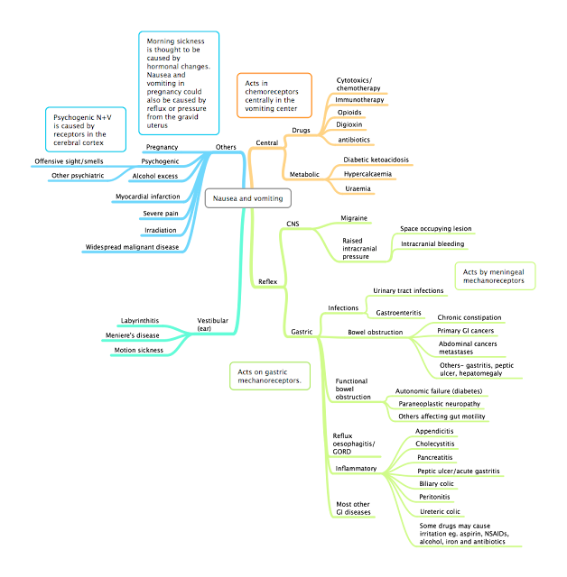
If a patient presents with nausea and vomiting, a clear cause should be established before perscribing antiemetics, as nausea and vomiting may be a sign of serious illness such as diabetic ketoacidosis, and may be reversed by treating the cause, for example in digoxin overdose. The cause of nausea and vomiting may be apparent with history and examination. patients with persistent nausea and vomiting should also be assessed for their fluid status, and for any electrolyte abnormalities caused by the vomiting. Causes of nausea and vomiting Nausea and vomiting may be caused centrally through stimulation of the vomiting center and the chemoreceptor trigger zone in the medulla, or caused afferent signals due to irritation in other areas of the body, usually in the gastrointestinal tract, but also due to disease in the central nervous system or the vestibular system in the ear. 

Lung Cancer

Lung cancer Definition : Uncontrolled invasive (malignant) growth originating from the lungs Presentation:   Depends on the site of the tumour and the type of cancer.  Patients may present with Chest signs and symptoms : (more common in cancers occurring centrally in the bronchial tree, late symptoms in peripheral cancers, may also present on a background of existing lung problems eg. COPD or emphysema)  *Haemoptysis *Cough Wheeze Stridor Pain Hoarse voice clubbing lymphadenopathy localized chest signs superior vena cava obstruction - caused by compression of the superior vena cava

Chemotherapy

Image
Malignancy occurs when cells mutate to replicate uncontrollably, outside of the body's normal control mechanisms and immune response, and gains the ability to invade into other tissues. Cell cycle As cancer cells tend to replicate faster than other cells, chemotherapy primarily affects cancer cells by affecting their replication.