Posts

Showing posts from April, 2014

Pericarditis ECG signs

Image
Immediate ECG changes (stage1)  Widespread, saddle shaped ST elevation (except AvR and lead 1)  PR interval depression (except AvR and lead 1)  Reciprocal changes in lead 1 and AvR.  Sinus tachycardia due to pain or effusion.  (taken from: http://cdn.lifeinthefastlane.com/wp-content/uploads/2011/03/V5-pericarditis.jpg)  1-3 weeks (stage 2)  Flattened T waves, no ST elevation or PR interval depression.  3- several weeks (stage 3)  Inverted T waves several weeks + (stage 4) ECG returns to normal *not all patients go through the later stages of ECG changes. http://lifeinthefastlane.com/ecg-library/basics/pericarditis/ (this is a brilliant site!) http://emedicine.medscape.com/article/156951-overview
Type 1 and Type 2 Diabetes

Image
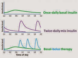
Diabetes is characterised by chronic hyperglycaemia caused by increased insulin resistance or reduced insulin release.  Aeitiology:  Diabetes may be primary or secondary. The main types of diabetes include type 1 diabetes, type 2 diabetes, secondary diabetes, and gestational diabetes. This post mainly discusses type 1 and type 2 diabetes. The main differences between type 1 and type 2 diabetes are summarised below:  Type 1 diabetes Type 2 diabetes Caused by the autoimmune destruction of B cells in the islet of Langerhans. Caused by increased peripheral insulin resistance and reduced insulin secretion. Tends to present before puberty, but may present at any age. Tends to occur in patients above 40 but increasingly affects younger individuals. Polygenetic, associated with inheritance of HLA types: HLA-DR3 and HLA-DR4. Associated with other autoimmune conditions. Polygenetic ...

Pulsus paradoxus

"Pulsus paradoxus" refers to an abnormal decrease of over 10mmHg in pulse pressure and subsequently pulse volume on inspiration. It is caused by a decrease in intrathoracic pressure.  On inspiration the intrathoracic pressure decreases The pressure intrathoracically is lower than the pressure extrathoracically which leads to an increased venous return to the right ventricles The right ventricles bulges into the left side reducing left ventricular volume.  Reduced intrathoracic pressure also leads to dialation of the pulmonary vessels Blood from the right ventricle pools in the lungs leading to reduced blood returning to the left ventricles Reduced blood in the left ventricles leads to a reduced stroke volume according to starling's law.  Reduced thoracic pressure on the aorta also contributes to the reduced pulse pressure Reduction of pulse pressure leads to a weaker or absent pulse volume being felt whist a contraction is heard on auscultation.  Pu...

Non pigmented skin cancers

Image
Non pigmented skin cancers (click to enlarge)

Pigmented lesions (Melanoma differentials)

Image
Differentials of pigmented lesions  (click to enlarge) - search on dermnetnz.org for more pictures! (also right click and open in new tab to see it in full size)  *correction - lentigo maligna melanoma is an invasive melanoma whereas lentigo maligna itself is a melanoma in situ. This tends to occur on the face in more elderly patients.

Skin anatomy

Image
Anatomy of the skin